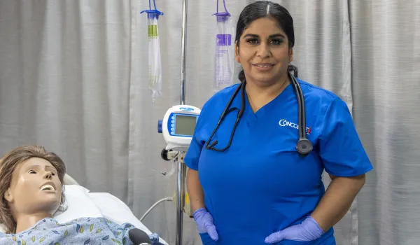
Concorde Staff

Sonography, or ultrasound imaging, is a noninvasive diagnostic tool that allows medical professionals to visualize internal organs and monitor health conditions. As the diagnostic imaging field continues to grow, so does the demand for skilled sonographers. There are several types of sonographers, each specializing in different areas of the body, such as the heart or muscles.
Understanding the different sonography specialties can provide valuable insight into the diverse career paths within this field. The U.S. Bureau of Labor Statistics projects an 11% growth in diagnostic and medical sonographer jobs from 2023 to 2033 , so there are promising opportunities for those entering the profession. Sonography is a critical, rewarding, and sought-after discipline that plays an important role in diagnosing and monitoring medical conditions such as cardiac care and prenatal health. This guide discusses the different types of sonographers and their career pathways.
What does a Sonographer do?
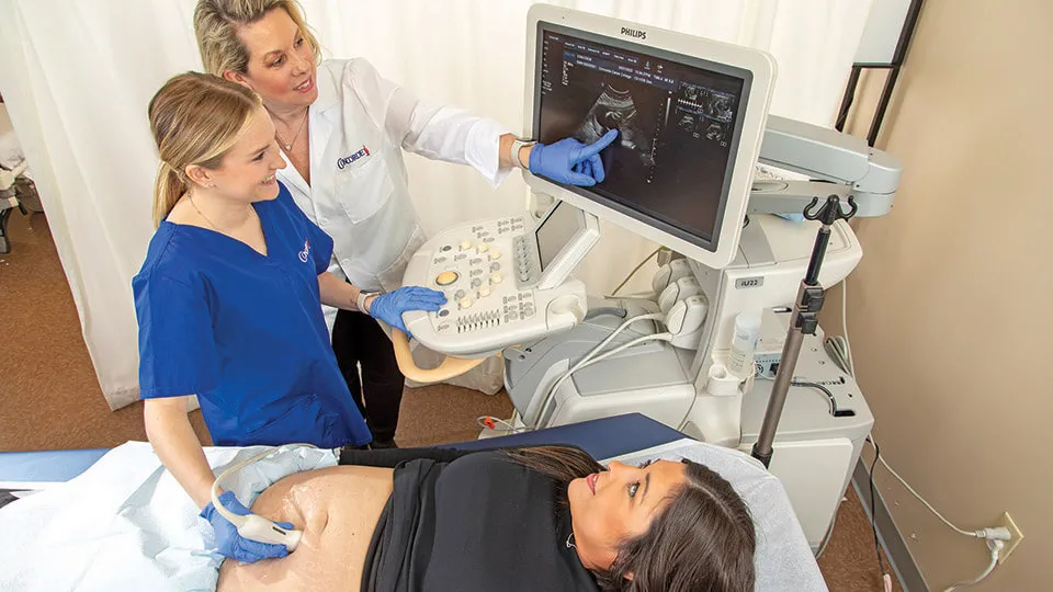
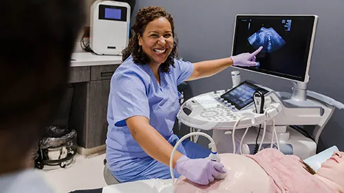
A sonographer is a health care professional who uses ultrasound technology to create images of the inside of the body, known as sonograms or ultrasound scans. These images help doctors diagnose and monitor medical conditions. These professionals typically specialize in different medical disciplines, such as cardiovascular, obstetric, gynecological, or musculoskeletal sonography. Sonographers operate ultrasound equipment that sends high-frequency sound waves into the body. The signals bounce off tissues and organs, creating real-time images that are displayed on a monitor.
These images assist physicians in diagnosing conditions such as tumors, heart diseases, pregnancies, and joint injuries. Sonographers must have technical knowledge of ultrasound equipment, a deep understanding of human anatomy, and the ability to interpret the images produced. They often work closely with doctors to ensure accurate diagnoses and contribute to patient care. They may work in hospitals, clinics, or private practices, depending on their specializations. A sonographer generally needs to complete formal education and training, typically earning an associate or bachelor's degree in diagnostic medical sonography. They may also need to obtain certification.
Related: Sonography Career Guide: Here's What To Know
1. Diagnostic Medical Sonographer
Diagnostic sonography is often considered a general role within the specialty. A diagnostic medical sonographer performs a variety of examinations, such as helping to diagnose gallstones or tumors with abdominal ultrasounds. A diagnostic medical sonographer must understand human anatomy and medical terminology, as their role requires accurately interpreting images for physicians. These sonographers typically work in hospitals, clinics, or private practices, collaborating with doctors and other health care professionals.
In addition to their technical skills, diagnostic medical sonographers need to interact effectively and explain procedures to make sure patients are comfortable during their procedures. Health care professionals entering this field typically need to hold an associate degree in diagnostic medical sonography and certification from recognized agencies. Concorde Career College offers its Diagnostic Medical Sonography Program at 11 campuses. This associate degree training program can be completed in as few as 20 months. The versatility of this role allows sonographers to experience diverse health care settings and achieve high levels of diagnostic accuracy.
Related: Ultrasound Technician vs Sonographer: What's the Difference
2. Cardiovascular Sonographer

A cardiovascular, echocardiographer, or cardiac sonographer specializes in imaging the heart and blood vessels using ultrasound technology. This role is critical in cardiac care, as these professionals perform echocardiograms to monitor heart function and conduct vascular studies to assess blood flow. Cardiovascular sonographers use advanced imaging equipment, including 2D and 3D technologies, to provide detailed insights into heart conditions such as valvular diseases or congenital issues.
Cardiovascular sonographers often work closely with cardiologists and other medical professionals to interpret images and provide important medical data for diagnosis and treatment. Specialized training and certification are required for this role, including expertise in understanding the complexities of the cardiovascular system. Due to the growing prevalence of heart disease around the world , the health care industry's demand for cardiovascular sonographers remains high. Concorde's offers its Cardiovascular Sonography Program at 12 campuses for students wanting to train for this career. This associate degree can be completed in as few as 20 months.
3. Obstetric and Gynecological Sonographer
An obstetric and gynecological sonographer plays an essential role in women's health. They specialize in imaging during pregnancy and assessing female reproductive health. These sonographers perform a variety of ultrasound examinations, such as monitoring fetal development during pregnancy and conducting pelvic exams to evaluate the health of a patient's reproductive organs.
Obstetric sonography has such a critical role in prenatal health care because these sonographers perform regular obstetric ultrasounds to track the growth and development of the fetus. Working alongside obstetricians and gynecologists, they help ensure that mothers and babies are healthy throughout pregnancies. This specialty requires a deep understanding of pregnancy milestones and women's health, so sonographers in this field must undergo specific training and certification. The frequency of obstetric ultrasounds, typically at least twice during pregnancy, makes this role particularly rewarding, as sonography directly contributes to the health and well-being of both mother and child.
Related: Ultrasound vs Radiology
4. Vascular Sonographer

A vascular sonographer focuses on imaging the circulatory system to help diagnose conditions relating to blood vessels. These health care professionals use specialized ultrasound technology, including Doppler imaging, to assess blood flow and detect conditions such as deep vein thrombosis, peripheral artery disease, and aneurysms. Vascular sonographers often collaborate with vascular surgeons and other specialists to provide critical diagnostic information for patients suffering from circulatory issues.
With the increasing incidence of stroke, aneurysms, and other vascular diseases, the demand for vascular sonographers has grown. These professionals are essential in identifying and monitoring circulatory conditions. They help doctors and other health care professionals make informed decisions regarding patient care. Special training and certification are required for this specialized field, ensuring sonographers are skilled in understanding vascular anatomy and performing detailed assessments.
5. Musculoskeletal Sonographer

A musculoskeletal sonographer specializes in imaging muscles, joints, and soft tissues. This field has seen growing demand due to its use in sports medicine and orthopedics. These sonographers perform dynamic imaging techniques to assess conditions such as tendon tears, joint inflammation, or muscle injuries. They work closely with orthopedic specialists and sports medicine professionals to diagnose and monitor musculoskeletal conditions, providing vital information for effective treatment plans.
Musculoskeletal sonography is also emerging in rheumatology, where it helps track conditions, such as rheumatoid arthritis, by detecting inflammation in the joints. The sonography role is becoming increasingly important in trauma assessment and long-term joint health. Specialized training and certifications are required to ensure sonographers are equipped with the necessary skills to perform intricate, accurate imaging.
Related: Is Health Care Career Training Right for You?
Preparing for a Successful Career as a Sonographer
Sonography represents a diverse and growing field within health care, with a unique focus and specialization. Sonographers play a major role in patient care, helping to diagnose cardiovascular conditions, monitor fetal development, and assess musculoskeletal injuries. As technology continues to advance and telemedicine grows, the demand for these health care professionals is projected to remain high.
Sonography is a field with strong job prospects and opportunities for specialized training and certification. Though the job outlook is promising, career outcomes can depend on specific training, hands-on experience, and chosen specialty. Concorde offers diagnostic medical sonography and cardiovascular sonography programs, both of which can be completed in just 20 months. Take the first step toward your sonographer training by contacting an admissions representative.
Related: Earn an Associate of Science in Diagnostic Medical Sonography
Frequently Asked Questions
Here are some frequently asked questions:
What are the latest advancements in diagnostic medical sonography technology?
The latest diagnostic medical sonography technology includes 3D and 4D imaging, elastography, and wearable ultrasound devices.
What are the most common cardiovascular conditions diagnosed through sonography?
Sonography can diagnose many cardiovascular conditions, such as heart valve abnormalities, coronary artery disease, and congenital heart defects.
How has 3D and 4D imaging technology impacted cardiovascular sonography?
3D echocardiography offers a more detailed view of the patient's heart, while 4D echocardiography allows sonographers a view in real time. This has the potential to speefd up diagnoses and treatment.
What are the current trends in fetal imaging techniques for obstetric sonography?
Current trends in fetal imaging include 3D and 4D ultrasound, elastography, and volumetric ultrasound. These techniques help improve diagnostic capabilities and provide more detailed images of the fetus.
How do gynecological sonographers contribute to early detection of reproductive health issues?
An ultrasound offers a clear view of the uterus, cervix, ovaries, and fallopian tubes. It can detect ovarian cysts, uterine fibroids, and endometriosis. This helps with early detection and improves treatment outcomes.
How does vascular sonography aid in the diagnosis and treatment of peripheral artery disease?
Vascular ultrasound, a non-invasive imaging test, helps diagnose and treat peripheral artery disease (PAD) by creating pictures of blood flow in the arteries.
What are the emerging applications of musculoskeletal sonography in sports medicine?
A musculoskeletal ultrasound is a non-invasive scan that is often used in sports medicine. It can help diagnose and monitor injuries, assess muscle imbalances, and support rehabilitation.
How is musculoskeletal sonography being used in rheumatology and orthopedics?
Musculoskeletal sonography is used to inspect soft tissues like tendons, ligaments, muscles, and joints. This helps assess inflammatory changes, fluid collections, bone erosions, and guiding procedures like injections. It's also helpful for diagnosing and monitoring conditions like arthritis, tendinitis, and ligament injuries.
What are the current certification requirements for different types of sonographers?
To become a certified sonographer in the USA, students need to complete an accredited program, usually an associate's degree, and pass an exam by the American Registry for Diagnostic Medical Sonography (ARDMS) or Cardiovascular Credentialing International (CCI). The exact requirements depend on the specialty, including areas like obstetrics or gynecology, vascular technology, abdominal imaging, musculoskeletal, or echocardiography.
Diagnostic Medical Sonographers and Cardiovascular Technologists and Technicians, Including Vascular Technologists : Occupational Outlook Handbook: : U.S. Bureau of Labor Statistics. (2019, April 12). Bls.gov; U.S. Bureau of Labor Statistics. https://www.bls.gov/ooh/healthcare/diagnostic-medical-sonographers.htm
News Medical. (2025, January 27). News-Medical. News-Medical. https://www.news-medical.net/news/20250127/Alarming-cardiovascular-disease-statistics-highlighted-in-2025-report.aspx
Take The Next Step Towards a Brighter Future
Interested in learning more about our Cardiovascular Sonography program?
We have a Concorde representative ready to talk about what matters most to you. Get answers about start dates, curriculum, financial aid, scholarships and more!
Related Articles
View All


Latest Articles
View All